
NMC CBME & MUHS Compliant Peer-Reviewed Clinical Vault • GMC Chhatrapati Sambhajinagar

Tag: general pathology

  • Pathology Notes MBBS 2nd Year: High-Yield Summary & How to Make Your Own

    ✅ Clinically Verified MBBS Resource • Written by Shrikant Bhosale (GMC Aurangabad) • NMC CBME Aligned

    Pathology is the bridge subject of MBBS. It explains the mechanism behind every clinical sign you will meet on the wards, and it carries serious weight in university exams and NEET PG. The students who struggle are almost never the ones who lack notes — they are the ones using someone else’s notes they never understood.

    This is a high-yield map of second-year pathology and, more importantly, how to build notes you actually own. All the material below is written from scratch; never rely on pirated textbook PDFs.

    What second-year pathology actually covers

    General pathology

    • Cell injury, cell death and adaptations — reversible vs irreversible injury, necrosis vs apoptosis, hypertrophy, hyperplasia, metaplasia, dysplasia.
    • Inflammation and repair — acute and chronic inflammation, mediators, granulomas, wound healing, regeneration and fibrosis.
    • Hemodynamic disorders — oedema, thrombosis, embolism, infarction, shock.
    • Neoplasia — nomenclature, benign vs malignant, carcinogenesis, tumour markers, staging and grading.
    • Immunopathology — hypersensitivity, autoimmunity, immunodeficiency, transplant rejection.
    • Genetics — mutations, chromosomal disorders, inheritance patterns.
    • Haematology — anaemias, leukaemias, lymphomas, coagulation.
    • Environmental and nutritional pathology.

    Systemic pathology

    Organ-based pathology across cardiovascular, respiratory, gastrointestinal, hepatobiliary, renal, endocrine, musculoskeletal, nervous system and skin — the same general mechanisms applied to organs.

    The note structure that works

    For every disease, use the same skeleton so your brain can compare across conditions:

    1. Definition — one line.
    2. Etiology / risk factors.
    3. Pathogenesis — the mechanism, in a flow of arrows.
    4. Morphology — gross and microscopic.
    5. Clinical features.
    6. Investigations.
    7. Complications and prognosis.

    Once every topic lives in the same seven boxes, revision becomes navigation rather than re-reading.

    Make notes that actually work

    • Draw, don’t just write. Flowcharts for pathogenesis, simple diagrams for morphology.
    • Compare, don’t list. A table of “necrosis vs apoptosis” teaches more than two paragraphs.
    • Link to clinics. Write one clinical example beside each mechanism.
    • Keep them short. If a page is dense with prose, it is a book, not a note.
    • Make them yourself. The act of condensing is the learning.

    How to study pathology in second year

    1. Read the topic from your main textbook, once, actively.
    2. Condense it into the seven-box structure above.
    3. Add 5–10 flashcards (mechanisms and morphology) using active recall and spaced repetition.
    4. Solve pathology previous-year questions on that topic the same week.
    5. Revisit on a spaced schedule — 3 days, then 10 days, then a month.

    High-yield topics that recur

    • Necrosis types and apoptosis pathways.
    • Acute inflammation mediators and chronic granulomatous inflammation.
    • Wound healing and factors affecting it.
    • Thrombosis, embolism and infarction.
    • Carcinogenesis, tumour markers, staging vs grading.
    • Anaemia classification and leukaemia/lymphoma basics.
    • Nephrotic vs nephritic syndromes.
    • Myocardial infarction pathology and the timeline of changes.

    Frequently asked questions

    How do I make pathology notes for MBBS 2nd year?

    Use one fixed skeleton for every disease — definition, etiology, pathogenesis, morphology, clinical features, investigations and complications — and draw flowcharts for mechanisms. Keep each topic short and rely on your own condensing rather than copied text.

    Is pathology hard in MBBS 2nd year?

    It is voluminous rather than conceptually hard. Because it repeats the same mechanisms across many organs, students who learn general pathology properly find systemic pathology far easier.

    Does pathology matter for NEET PG?

    Yes. Pathology is one of the highest-yield subjects in NEET PG and INI-CET, so notes built properly in second year pay off directly at the PG entrance stage.

    Can I use ready-made pathology notes?

    You can use them as a supplement, but only notes you make yourself force the retrieval that builds memory. Pirated textbook PDFs are also a copyright and ethical problem — avoid them.

    Build your reading habit from the first-year book list, and learn the method that makes notes stick with active recall and spaced repetition.
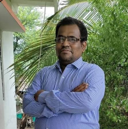
    Shrikant Bhosale — Final Year MBBS Student

    Written by Shrikant Bhosale

    Final Year MBBS Student at Government Medical College, Chhatrapati Sambhajinagar (Aurangabad), Maharashtra. I write the study, ward and career guides I wish existed when I started MBBS — grounded in the real NMC curriculum, real university exams and real ward experience. More about me →

    Educational use only. This guide is written for medical students and is not medical advice. Clinical protocols, drug doses and guidelines change — always verify against the latest standard textbooks and official NMC / institutional guidance before applying anything in practice. Read the full disclaimer.

    📌 Executive Summary & High-Yield Takeaways

    • Pathology is the bridge between preclinical sciences and clinical medicine — master the ‘etiopathogenesis’ framework.
    • The 6-point answer structure for every disease: Definition -> Etiology -> Pathogenesis -> Gross morphology -> Microscopic morphology -> Clinical features / Complications.
    • Neoplasia and Cell Injury: High-yield foundation chapters; 40% of general pathology exam questions come from here.
    • Learn microscopy diagrams: Drawing neat, colored (H&E pink and blue) sketches secures top marks in practical exams.

    Frequently Asked Questions (FAQ)

    Common questions asked by medical students regarding this topic:

    Q: What is the best way to write a 10-mark pathology question in exams?

    Follow the standardized framework: Definition, Etiology & Risk Factors, Pathogenesis flowchart, Gross appearance (size, shape, color, cut section), Microscopic features (H&E diagram), and Clinical Course.

    Q: Which chapters in Robbins are highest yield for 2nd year MBBS?

    Cell Injury & Adaptation, Inflammation & Repair, Neoplasia, Hemodynamic Disorders, Atherosclerosis & Ischemic Heart Disease, Cirrhosis, and Glomerulonephritis.

    Q: How should I prepare for pathology practical slide viva?

    Practice identifying key morphological hallmarks (e.g., Reed-Sternberg cells in Hodgkin lymphoma, granulomas in tuberculosis, signet ring cells in gastric adenocarcinoma) and draw labeled pencil sketches in your record book.

    Shrikant Bhosale, Final Year MBBS Student

    Written by Shrikant Bhosale

    Final-Year MBBS Student at Government Medical College, Chhatrapati Sambhajinagar (Aurangabad), Maharashtra. Affiliated with Maharashtra University of Health Sciences (MUHS). Written to provide unbloated, peer-tested medical education resources grounded in real hospital ward practice and NMC CBME university examinations.

    View Author Profile & Clinical Postings • Editorial Standards • Submit Errata

  • General Pathology Notes: Free High-Yield Flowcharts & Summaries

    ✅ Clinically Verified MBBS Resource • Written by Shrikant Bhosale (GMC Aurangabad) • NMC CBME Aligned

    General pathology is the foundation of everything else in pathology, and it is the part most worth learning properly because it repeats across every organ system. These are original, plain-language summaries and flowcharts to make revision fast — no scraped textbook text, no pirated PDFs.

    Use this page as your revision map, then build your own full notes using the system in my pathology notes guide.

    Cell injury and cell death

    Reversible injury → cell swelling, fatty change, blebbing. Irreversible injury → membrane damage, mitochondrial failure, calcium influx, enzymatic digestion.

    Mechanisms to remember as a chain:

    Ischaemia → ATP depletion → failure of Na⁺/K⁺ ATPase → cell swelling → anaerobic metabolism → acidosis → ribosome detachment → decreased protein synthesis → mitochondrial damage → irreversible injury → necrosis or apoptosis.

    Know the difference cold: necrosis (pathological, inflammatory, membrane rupture) versus apoptosis (regulated, energy-dependent, no inflammation), and the subtypes of necrosis — coagulative, liquefactive, caseous, fat and fibrinoid.

    Adaptations

    • Hypertrophy — increase in cell size.
    • Hyperplasia — increase in cell number.
    • Atrophy — decrease in size and function.
    • Metaplasia — reversible change of one adult cell type to another.
    • Dysplasia — disordered growth, a precursor to malignancy.

    Acute inflammation

    The five cardinal signs: redness, heat, swelling, pain and loss of function.

    • Vascular phase: vasodilation, increased permeability.
    • Cellular phase: margination, rolling, adhesion, transmigration, chemotaxis, phagocytosis.
    • Mediators: histamine, prostaglandins, leukotrienes, cytokines, complement, nitric oxide.

    Then know the morphological patterns — serous, fibrinous, suppurative, ulcerative — and the outcomes: resolution, healing by fibrosis, chronic inflammation or abscess.

    Chronic inflammation and granulomas

    Characterised by mononuclear infiltration, tissue destruction and repair. Granuloma = collection of activated macrophages (epithelioid cells, giant cells) — caseating (tuberculosis) or non-caseating (sarcoidosis, Crohn’s, foreign body).

    Haemodynamic disorders

    • Oedema: increased hydrostatic pressure, reduced oncotic pressure, lymphatic obstruction, sodium retention.
    • Thrombosis: Virchow’s triad — endothelial injury, stasis/turbulence, hypercoagulability.
    • Embolism: thromboembolism, fat, air, amniotic fluid.
    • Infarction: red vs white; lung, liver and kidney pattern differences.
    • Shock: hypovolaemic, cardiogenic, distributive (septic, anaphylactic, neurogenic), obstructive.

    Neoplasia

    • Benign vs malignant: differentiation, rate of growth, local invasion, metastasis.
    • Carcinogenesis: initiation → promotion → progression, with oncogenes and tumour suppressor genes (RB, p53).
    • Spread: direct, lymphatic, haematogenous, transcoelomic.
    • Staging vs grading: grading = differentiation; staging = extent (TNM).
    • Tumour markers: learn the classic associations (e.g., AFP, CEA, CA-125, PSA).

    How to revise general pathology fast

    1. Redraw each flowchart from memory daily for a week.
    2. Tabulate every “versus” pair the examiner loves: reversible vs irreversible, necrosis vs apoptosis, benign vs malignant, transudate vs exudate.
    3. Attach one clinical example to each mechanism.
    4. Solve previous-year questions topic by topic.

    Frequently asked questions

    Where can I get free general pathology notes?

    Use original summaries and flowcharts like those on this page, and build your own structured notes from your prescribed textbook. Avoid pirated textbook PDFs, which are both unethical and often unreliable.

    What are the most important topics in general pathology?

    Cell injury and death, inflammation and repair, haemodynamic disorders, neoplasia and immunopathology — these underpin the entire subject and dominate exam questions.

    How do I remember pathology flowcharts?

    Redraw them from memory at increasing intervals; the act of reconstructing the chain is far more effective than re-reading it. Pair this with the flashcards from your general pathology topics.

    Are these notes enough for MBBS exams?

    They are a revision framework, not a substitute for your textbook. Use them to structure and test your knowledge, and expand each point from your prescribed reading.

    Shrikant Bhosale — Final Year MBBS Student

    Written by Shrikant Bhosale

    Final Year MBBS Student at Government Medical College, Chhatrapati Sambhajinagar (Aurangabad), Maharashtra. I write the study, ward and career guides I wish existed when I started MBBS — grounded in the real NMC curriculum, real university exams and real ward experience. More about me →

    Educational use only. This guide is written for medical students and is not medical advice. Clinical protocols, drug doses and guidelines change — always verify against the latest standard textbooks and official NMC / institutional guidance before applying anything in practice. Read the full disclaimer.

    📌 Executive Summary & High-Yield Takeaways

    • Cellular adaptations: Hypertrophy, Hyperplasia, Atrophy, Metaplasia — memorize real clinical examples for each.
    • Necrosis vs Apoptosis: Essential comparison table tested in every university exam.
    • Chemical mediators of inflammation: Histamine, Prostaglandins, Leukotrienes, Cytokines (TNF, IL-1), and Complement cascades.
    • Carcinogenesis multistep model: Initiation, Promotion, Progression; Proto-oncogenes vs Tumor Suppressor genes (p53, RB).

    Frequently Asked Questions (FAQ)

    Common questions asked by medical students regarding this topic:

    Q: What are the key differences between necrosis and apoptosis?

    Necrosis is always pathological, involves cell swelling, plasma membrane disruption, enzymatic digestion, and elicits an inflammatory reaction. Apoptosis can be physiological or pathological, involves cell shrinkage, chromatin condensation, apoptotic body formation, and elicits no inflammation.

    Q: What is the role of p53 as the ‘guardian of the genome’?

    p53 is a tumor suppressor protein that senses DNA damage. It halts the cell cycle in G1 phase via p21 to allow DNA repair; if damage is irreparable, p53 upregulates BAX and triggers apoptosis.

    Q: What are the cardinal signs of acute inflammation?

    The five cardinal signs (Celsus & Virchow) are Rubor (redness), Calor (heat), Tumor (swelling), Dolor (pain), and Functio laesa (loss of function).

    Shrikant Bhosale, Final Year MBBS Student

    Written by Shrikant Bhosale

    Final-Year MBBS Student at Government Medical College, Chhatrapati Sambhajinagar (Aurangabad), Maharashtra. Affiliated with Maharashtra University of Health Sciences (MUHS). Written to provide unbloated, peer-tested medical education resources grounded in real hospital ward practice and NMC CBME university examinations.

    View Author Profile & Clinical Postings • Editorial Standards • Submit Errata